Полезное:
Как сделать разговор полезным и приятным
Как сделать объемную звезду своими руками
Как сделать то, что делать не хочется?
Как сделать погремушку
Как сделать так чтобы женщины сами знакомились с вами
Как сделать идею коммерческой
Как сделать хорошую растяжку ног?
Как сделать наш разум здоровым?
Как сделать, чтобы люди обманывали меньше
Вопрос 4. Как сделать так, чтобы вас уважали и ценили?
Как сделать лучше себе и другим людям
Как сделать свидание интересным?

Категории:
АрхитектураАстрономияБиологияГеографияГеологияИнформатикаИскусствоИсторияКулинарияКультураМаркетингМатематикаМедицинаМенеджментОхрана трудаПравоПроизводствоПсихологияРелигияСоциологияСпортТехникаФизикаФилософияХимияЭкологияЭкономикаЭлектроника
Chromosome structure
Fluorescent staining highlights the chromosomes of the bacterial pathogen Salmonella enteriditis. The cytoplasm is orange, and the chromosome fluoresces in bright yellow. Some bacteria appear to have more than one chromosome because they are in the process of dividing.
DNA is associated with proteins; each DNA with its associated proteins is called a chromosome. Chromosome consists of two sister chromatids, which contain densely organized double helix of DNA, and centromere (primary constriction) that connects them. This organization holds true for prokaryotic and eukaryotic cells and even for viruses.
Packaging of the DNA into chromosomes serves several important functions.
· The chromosome is a compact form of the DNA that readily fits inside the cell.
· Packaging the DNA into chromosomes serves to protect the DNA from damage. Completely naked DNA molecules are relatively unstable in cells. In contrast, chromosomal DNA is extremely stable, allowing the information encoded by the DNA to be reliably passed on.
· Only DNA packaged into a chromosome can be transmitted efficiently to both daughter cells each time a cell divides.
· The chromosome gives an overall organization to each DNA molecule. This organization facilitates gene expression as well as the recombination between parental chromosomes that generates the diversity observed among different individuals of any organism.
Chromosomal DNA contains genes, regulatory elements and additional nucleotide sequences. All eukaryotic cells have multiple linear chromosomes; the number of chromosomes typically varies from 2 to less than 50. An average human chromosome is about 45 nm in length of 130·106 bp. If the DNA helix of a diploid human cell rolls out, its length would have been about 2 meters. During mitosis at metaphase and prometaphase chromosomes have a length of from 2 to 11 microns, which enables microscopic examination of stained individual chromosomes. Chromosomes are located in the nuclei of all human cells (except mature red blood cells, they do not have nuclei), and each cell contains 23 pairs of different chromosomes. The number, size, and shape of chromosomes are clearly defined and specific to each species.
Half of the molecular mass of a eukaryotic chromosome is protein. In eukaryotic cell DNA with its associated proteins is called chromatin and the majority of the associated proteins are small, basic proteins called histones. Non-histone proteins are associated with the chromosome too. These proteins include the numerous DNA-binding proteins that regulate the transcription, replication, repairing, and recombination of cellular DNA. Most compaction in eukaryotic cells is the result of the regular association of DNA with histones to form structures called nucleosomes.
Prokaryotic cells typically have smaller genomes, but need to compact their DNA. E. coli must pack its approximately 1 mm chromosome into a cell that is only 1 μm in length. Bacteria have no histones and nucleosomes, but they have other small basic proteins that may serve similar functions. Prokaryotic cells typically have only one complete copy of their chromosome(s) that is packaged into a structure called the nucleoid. The most studied prokaryotes (e.g. E. coli, B. subtilis) have single circular chromosomes; but there are numerous prokaryotic cells with multiple chromosomes, linear chromosomes, or even both. Prokaryotes also frequently carry one or more smaller independent circular DNAs, called plasmids. Unlike the chromosomal DNA, plasmids typically are not essential for bacterial growth. They carry genes that confer desirable traits to the bacteria, such as antibiotic resistance. Also distinct from chromosomal DNA, plasmids can be present in many complete copies per cell.
The majority of eukaryotic cells are diploid; that is, they contain two copies of each chromosome. The two copies of a given chromosome are called homologs; one is derived from each parent. Not all cells in a eukaryotic organism are diploid; some eukaryotic cells are either haploid or polyploid. Haploid cells contain a single copy of each chromosome and are involved in sexual reproduction (for example, sperm and eggs are haploid cells). Polyploid cells have more than two copies of each chromosome, e.g. hepatocytes of mammals.
3. Central dogma of molecular biology
Chromosomal DNA function is to be the template for RNA molecules synthesis, those subsequently move to the cytoplasm, where they determine the arrangement of amino acids within proteins. In 1956, Francis Crick referred to this pathway for the flow of genetic information as the central dogma. DNA is the template for its self-replication. RNA synthesis (transcription) is directed by a DNA template. The synthesis of proteins (translation) is directed by an RNA template. Most importantly, the last two processes are unidirectional; that is, RNA sequences are never determined by protein templates, nor DNA ever to be made on RNA templates.
Now it’s considered that proteins never serve as templates for RNA synthesis. However, RNA chains sometimes do act as templates for DNA chains of complementary sequence. Such reversals of the normal flow of information are very rare events compared with the enormous number of RNA molecules made on DNA templates. The central dogma as originally proclaimed approximately 60 years ago still remains essentially valid.
4. Cytogenetics as the science of chromosomes
Cytogenetics explores the structure and function of individual chromosomes or chromosome set as a whole.
It is believed that the beginning of cytogenetics as a science of the chromosome was in 1882, when Walter Fleming, cytologist and professor of anatomy, published the first illustration of chromosomes. However, back in 1880, he first used the term “mitosis” and named the contents of stained nucleus “chromatin”. Scientist has shown that during mitosis, after longitudinal chromosomes division, each half goes to one of the daughter cells.
In 1883 Е. van Beneden confirmed that the offspring chromosomes derived equally from both parents (due to the merger слияние of germ cells), their structure repeats maternal chromosome.
In 1888 German anatomist H.W.G. Waldeyer introduced the term “chromosome” to refer stained filamentous structures that were visible in cell during mitosis. In 1900 W.Sutton in U.S. and T.Boveri in Germany independently suggested that genes are located in chromosomes. This idea formed the basis for the chromosome theory of heredity. Combining such subjects as cytology and genetics, W.Sutton introduced the term “cytogenetics”.
In the late 19th and early 20th century cytogenetics investigated the exact number of chromosomes and sex dependence on the configuration of the sex chromosomes. Painter suggested hypothesis about sex dependence of X and Y chromosome configuration.
Only in 1956 D.Tijo and A.Levan determined that the diploid number of human chromosomes is 2n = 46, in the same year, this was confirmed by Ford and Hamerton.
In 1970 methods for the differential staining of chromosomes were developed. It made possible to identify finally all human chromosomes.
Cytogenetic methods (method of chromosome analysis) are based on microscopic examination of chromosomes structure and number.
The study of human chromosomes plays an important role in the diagnosis, prognosis and monitoring of treatment effectiveness not only in medical genetics but also in pediatrics, hematology, oncology, endocrinology and others.
5. General plan of prokaryotic and eukaryotic cell
Bacteria are prokaryotes, e.g. E.coli
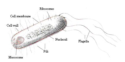
Structure of the bacteria E.coli
Eukaryotic cells
6. The unique structure of mitochondria
Mitochondria (along with chloroplasts) are unique among organelles in that they divide independently of the cell, contain circular strands of DNA, and have procaryotic-sized 70S ribosomes. These findings have prompted some intriguing speculations on their evolutionary origins
7. The main differences of prokaryotic and eukaryotic cell structure
Date: 2015-09-02; view: 436; Нарушение авторских прав; Помощь в написании работы --> СЮДА... |